 43
43

3D breast imaging improves accuracy
YOUNGSTOWN
Dr. Jonah Moon uses a loaf of bread analogy when describing how impactful 3D imaging has become to breast cancer screening.
Take a loaf of cinnamon raisin bread, for instance. When you do the 2-D mammogram, he said, you make out a bunch of raisins and stripes of cinnamon, but those raisins can be overlapping and hide something underneath.
But when you do the 3-D mammogram you can more carefully analyze what’s inside the bread, slice by slice. And at that time, he said, you may notice a stray walnut.
“Finding that walnut is like finding cancer hidden in the breast,” said Moon, the co-director of the Joanie Abdu Comprehensive Breast Care Center.
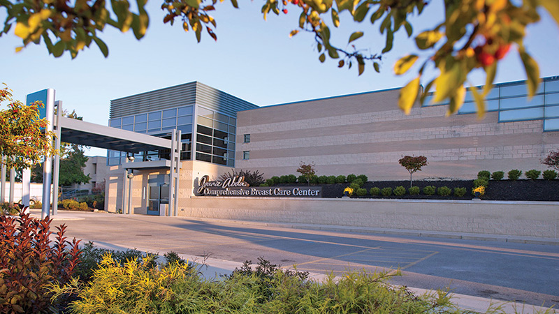
Mercy Health–Youngstown was the first to bring 3-D mammography to the Mahoning Valley in 2011 when it opened the Joanie Abdu Center. It’s now offered not only downtown, but at St. Joseph Hospital in Warren and the newly opened Poland Imaging Services, as well as its mobile mammovan unit.
“3-D is quickly becoming the standard of care,” Dr. Moon said.
What 3-D offers, he said, is the ability to see certain anomalies that just aren’t apparent on 2-D screens. Further, he said, sometimes a 2-D image will show something suspicious, but when looking at the image in 3-D it ends up being an overlap of tissue and not a cause for concern.
“A situation like that, creates a lot of anxiety for a woman who is called back for additional testing,” said Juli Dulay, manager of the Joanie Abdu Center. “She’s assuming it’s cancer, but it might be a non-cancerous or benign finding. Using 3-D technology dramatically reduces the number of false alarms.”
Dr. Moon and Dulay said standard 2-D digital scans are provided even when 3-D is used, giving physicians a complete picture of the breast architecture.
“3D definitely has a clinical advantage,” Dulay said. “The 3D scan is an additional sweep that takes 1-milimeter slice pictures through the breast. It opens the structure of the breast, allowing us to see more cancers.”
The 3D scan typically adds just one to two minutes to the screening.
National research, according to WebMd.com, shows cancer detection rates have improved by 40 percent when 3-D imaging is used in addition to 2-D. That’s because breast cancer outcomes notably improve, Dr. Moon said, the earlier the cancer is detected.
Downsides to 3-D breast imaging are minimal. Dr. Moon points out that 3-D scans do come with a greater radiation exposure than 2-D scans, about the same as a film X-ray, he said. The only time he doesn’t do 3-D scans is because of patient preference.
“The benefits definitely outweigh the downsides of it,” Dr. Moon said.
Dulay said the improvement from 2-D to 3-D is staggering; not unlike when we went from Kodak Instamatic to digital photography in personal picture taking.
“3-D is the newest technology available in mammography. It’s a huge clinical benefit and we have it readily available right here in the Mahoning Valley,” Dulay said. “It improves accuracy and overall visualization for the physicians, and provides greater peace of mind and reduced anxiety for patients.”